Քրոնիկական պոչուկացավը (կոկցիգոդինիան) բնորոշվում է պոչուկի հատվածում ավելի քան 6 ամիս տևող ցավով, որը սաստկանում է նստած դիրքում և/կամ ոտքի ելնելիս:
Թեև ախտորոշման հիմքում կլինիկական պատկերն է, անհրաժեշտ են լրացուցիչ պատկերային հետազոտություններ (մագնիսա-ռեզոնանսային շերտագրություն [ՄՌՇ] կամ նույնիսկ սցինտիգրաֆիա) բուժելի հիվանդությունների (ինչպիսիք են տվյալ շրջանի ուռուցքները) ախտանշանային ձևեր որոնելու համար: Այս քննությունները երբեմն օգնում են նաև պարզաբանել ցավի համար պատասխանատու ախտաֆիզիոլոգիական մեխանիզմները:
Պոչուկի դինամիկ ռենտգենագրությունը (նստած, կանգնած) էտալոնային հետազոտություն է: Դինամիկ գործողությամբ (ուղիղաղիքային մատնային զննում) պոչուկային ՈՒՁՀ-ն նորարարական այլընտրանք է, որը դեռևս լիովին չի վավերացվել:
Քրոնիկական պոչուկացավի ամենատարածված պատճառներն են գերշարժուն կամ կարծրացած պոչուկը և պոչուկային փուշը (սպիկուլան): Հաճախ բացահայտվում է վնասվածքային կամ հետծննդաբերական ծագում։
Սոցիալական և ցավային հետևանքներն այնպիսին են, որ արդարացնում են բուժօգնության դիմելը. նախ իրականացվում են մանուալ բուժումներ, ապա՝ ինֆիլտրացիա ու, անհրաժեշտության դեպքում, տեղային այլ միջոցների կիրառություն, և, ի վերջո, ամենից կայուն դեպքերի ժամանակ՝ վիրահատություն։
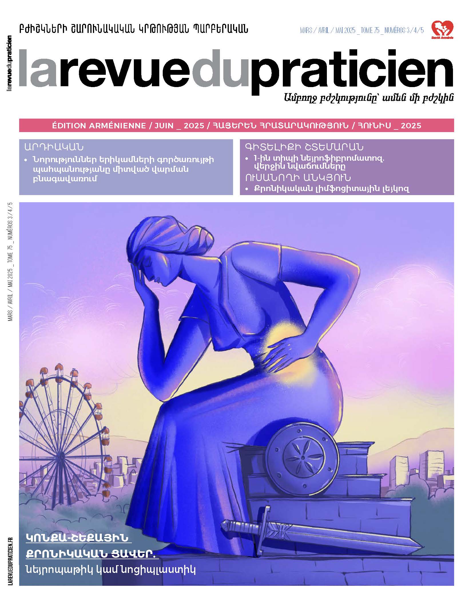
La coccygodynie chronique est définie par une douleur du c occyx évoluant depuis plus de six mois se majorant en position assise et/ou au relever. Si le diagnostic est avant tout clinique, des examens complémentaires d’imagerie (imagerie par résonance magnétique [IRM], voire scintigraphie) sont nécessaires pour rechercher des formes symptomatiques de pathologies curables telles que les tumeurs de la région. Ces examens permettent aussi parfois d’élucider les mécanismes physiopathologiques responsables des douleurs. Les radiographies dynamiques (assis, debout) du coccyx restent l’examen de référence. L’échographie coccygienne avec manoeuvre dynamique (toucher rectal) représente une alternative novatrice non encore complètement validée. Les causes les plus fréquentes de coccygodynie chronique sont les coccyx hypermobiles, rigides et les spicules coccygiens. On met souvent en évidence une origine traumatique ou post-obstétricale. Les conséquences sociales et douloureuses sont telles qu’elles justifient le recours aux soins : d’abord thérapies manuelles, puis infiltration et éventuellement utilisation de topiques, et enfin chirurgie pour les cas les plus rebelles.
Chronic coccygodynia is defined as pain in the c occyx which has been present for more than six months and which is aggravated by sitting down and/or rising. Although the diagnosis is primarily clinical, additional imaging examinations (MRI, even scintigraphy) are necessary to look for symptomatic forms of curable pathologies such as tumors in the region. These investigations can also help to elucidate the pathophysiological mechanisms responsible for the pain. Dynamic X-rays (sitting and standing) of the coccyx remain the reference exam. Coccygeal ultrasound with a dynamic maneuver (digital intrarectal mobilization of the coccyx) represents an innovative alternative that has not yet been fully validated. The most frequent causes are hypermobile or rigid coccyxes and coccygeal spicules. Traumatic or post-obstetric causes are often found. The social and painful consequences are such as justifying recourse to treatment. Firstly, manual therapies, then infiltrations and possibly the use of topicals, and finally surgery for the most intractable cases.